Home » Cartilage defects

Cartilage Defects: What They Mean, Why They Hurt, and How I Evaluate Them

Cartilage defects are a common finding in MRI reports, especially around the knee, but the wording can sound more alarming than the actual problem. Cartilage is the smooth, protective layer that covers the ends of bones inside a joint. It allows the joint to move with low friction and to bear load smoothly. When that surface is damaged, patients may develop pain, swelling, stiffness, catching, or a sense that the joint is not working normally.

In my practice, I often see Bangladeshi patients who read the word “defect” and immediately assume the joint is ruined. That is not always true. A cartilage defect can be small, moderate, or extensive; stable or unstable; isolated or part of a larger joint problem. The right treatment depends on the cause, the size, the location, and the patient’s symptoms.

What is a cartilage defect?

A cartilage defect means that part of the normal smooth joint surface has been injured, worn away, or lost. In some cases, only the cartilage is affected. In other cases, the underlying bone is also involved, and the report may describe an osteochondral lesion.

The knee is the joint where I most often discuss this problem, but cartilage defects can also occur in the ankle, hip, shoulder, and other joints. One important point I explain to patients is that cartilage has limited natural healing ability compared with skin or muscle. That is why some defects settle down with treatment while others continue to cause trouble over time.

Common causes of cartilage damage

There is no single cause for every patient. In Bangladesh, I commonly see cartilage damage after:

Sudden injury

A twisting injury, fall, sports trauma, or dislocation can directly damage the cartilage surface.

Repeated overload

Long-term stress from squatting, stair climbing, kneeling, heavy lifting, running, jumping, or frequent impact can gradually wear the joint surface.

Instability or malalignment

If a joint is unstable or the alignment is poor, force is distributed unevenly and cartilage can break down faster.

Meniscus or ligament injury

Damage to the meniscus or ligaments, especially around the knee, can increase pressure on the cartilage and accelerate wear.

Early degenerative change

In some older adults, cartilage defects are part of early osteoarthritis rather than a single injury.

The cause matters because treatment should address the real mechanism, not just the MRI wording.

Symptoms I ask about

Symptoms vary depending on the size of the defect, the joint involved, and whether other structures are injured. Patients may describe:

  • pain during walking, squatting, stairs, or sports
  • swelling after activity
  • stiffness, especially after rest
  • clicking or catching
  • locking in some cases
  • a feeling that the joint may give way
  • reduced confidence in using the joint

Small defects can be deceptive. A patient may feel only mild discomfort at first, then notice repeated swelling or pain when activity increases. Others come to clinic only after the joint starts interfering with work, prayers that involve kneeling, household activity, driving, or sport.

How I evaluate a cartilage defect

When I assess a patient, I do not rely on the MRI alone. I start with a careful history and examination.

History

I want to know whether the problem began after an injury, whether pain is constant or activity-related, whether there is swelling, and whether the patient has mechanical symptoms such as catching or locking.

Examination

I check swelling, tenderness, range of motion, alignment, muscle strength, and joint stability. I also look for signs of meniscus injury, ligament injury, or arthritis.

Imaging

X-rays are useful for assessing alignment, joint space, arthritis, and related bone changes. MRI is often better for showing cartilage injury and associated soft-tissue damage. If symptoms and scan findings do not match, I trust the clinical picture more than the report alone.

That point matters. A dramatic MRI sentence does not always mean surgery is required.

Treatment options

Treatment depends on the patient, the lesion, and the level of symptoms. Some patients improve without surgery. Others need a procedure because the defect is unstable, painful, or associated with other structural damage.

Non-surgical treatment

For many patients, especially those with smaller or stable defects, I usually begin with conservative care. This may include:

  • activity modification
  • weight management when relevant
  • guided physiotherapy
  • muscle strengthening around the joint
  • pain control when appropriate
  • avoiding repetitive movements that flare symptoms

For knee cartilage problems, strengthening the quadriceps, hip muscles, and core support can improve joint mechanics and reduce stress on the damaged surface. Random exercise is not the same as a structured rehabilitation plan. The goal is to protect the joint while improving function, not simply to rest forever.

Surgical treatment

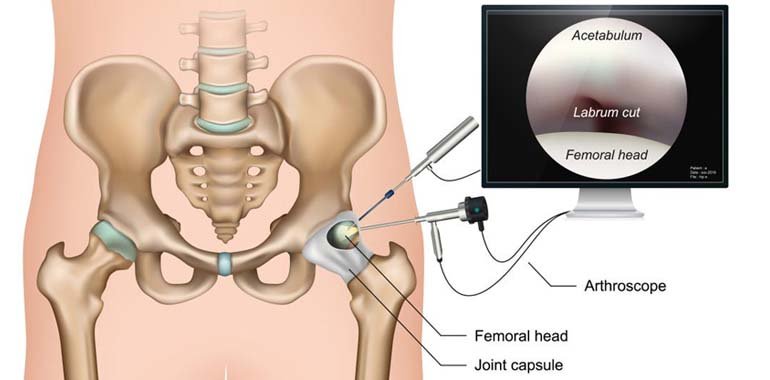
If pain, swelling, or mechanical symptoms continue despite proper conservative care, I may discuss arthroscopy or other cartilage procedures. Depending on the lesion, options can include cleaning unstable tissue, microfracture in selected cases, fixation of a fragment, or cartilage-restoration procedures.

The decision is individualized. Not every cartilage defect needs an operation, and not every lesion is suitable for the same procedure. Age, activity level, alignment, stability, and associated injuries all influence the plan.

When delay becomes a problem

I have seen many patients wait too long because they hope the pain will settle on its own or they want to avoid time away from work. That is understandable, especially in Bangladesh, where patients may be balancing family responsibilities, transport issues, and cost concerns. But repeated swelling, worsening pain, and continued overload can make the joint harder to manage later.

Knee Care by Dr. Md. Iftekharul Alam

Early evaluation does not automatically mean surgery. Often it simply means a better plan, clearer expectations, and a chance to prevent further damage.

Can a cartilage defect lead to arthritis?

It can increase the risk of future joint wear, especially if the underlying cause is not corrected. That does not mean every defect becomes arthritis, but it does mean persistent symptoms should not be ignored.

If the joint keeps swelling, catching, or giving way, the mechanics are not normal. In that setting, I advise a proper orthopedic review rather than repeated self-treatment.

Red flags that need urgent medical attention

Some symptoms deserve prompt assessment, especially after injury. Patients should seek urgent care if they have:

  • inability to bear weight
  • severe swelling after trauma
  • a locked joint that cannot move normally
  • major deformity after injury
  • fever with a hot, swollen joint
  • severe pain that is rapidly worsening
  • numbness, weakness, or loss of circulation symptoms

Those features may indicate a more serious injury, infection, or associated structural problem rather than a simple cartilage defect.

What patients in Dhaka and Bangladesh should remember

In my practice, I often explain that treatment has to fit real life. A patient may need to continue working, travel long distances, care for family members, or manage stairs at home. Because of that, the best plan is the one that balances symptom control, joint protection, and realistic rehabilitation.

That is why I focus on:

  • understanding the true cause
  • matching treatment to the lesion
  • avoiding unnecessary surgery
  • not dismissing persistent symptoms
  • building a recovery plan that the patient can actually follow

What this usually means for Bangladeshi patients

A cartilage defect does not always mean immediate surgery, but it does mean the joint deserves careful follow-up if pain, swelling, or catching continue. I usually discuss whether the plan should focus on load modification, rehabilitation, arthroscopy, or longer-term monitoring. The right decision depends on symptoms, size and location of the defect, and how the joint is functioning in daily life.

How I Match Symptoms to the Likely Problem

Pain, swelling, stiffness, locking, weakness, and instability do not all point to the same diagnosis. I usually relate the symptom pattern to age, injury history, weight-bearing pain, stair difficulty, squatting, sport demands, and night symptoms before deciding what is most likely.

For Bangladeshi patients, this early mapping is useful because it helps separate a problem that may respond to activity modification and physiotherapy from one that needs an X-ray, MRI, laboratory evaluation, or prompt orthopedic assessment.

When a cartilage problem needs more than rest

I usually become more concerned when cartilage-related pain is accompanied by swelling after activity, catching, repeated giving way, or loss of confidence in the joint. Those features suggest the issue may be affecting mechanics rather than causing only short-term irritation.

For Bangladeshi patients, the next step should be practical and diagnosis-driven. Some need physiotherapy and load adjustment, while others need MRI, arthroscopy planning, or treatment for associated ligament and meniscus problems.

A Practical Bangladesh Care Note

For patients in Bangladesh, access to imaging, physiotherapy, and follow-up can influence which treatment plan is most realistic. I usually discuss not only the diagnosis itself, but also whether the patient can return for review, continue exercises properly, and manage the demands of work, study, stairs, and long travel while recovering.

When cartilage damage needs closer evaluation

Cartilage injury can be difficult for patients to understand because the pain may seem out of proportion to the X-ray. In my practice, I consider the injury pattern, swelling, mechanical symptoms, prior trauma, and whether MRI is needed to define the defect more clearly. Some patients can be managed with strengthening and load control, while others need more targeted intervention.

The important point is that persistent swelling, locking, or repeated giving way should not be dismissed as simple wear and tear without proper assessment.

Related topics

When cartilage symptoms deserve a more focused workup

Cartilage injury often sounds vague to patients, but the pattern of pain, swelling, catching, and activity intolerance can be quite specific. I usually try to determine whether the problem is isolated cartilage damage, an unstable fragment, early arthritis, or a combined injury involving the meniscus or ligament structures.
That distinction is important in Bangladesh because treatment planning changes significantly depending on the exact pattern of damage.

References

  1. AAOS OrthoInfo: Articular Cartilage Restoration
  2. AAOS OrthoInfo: Knee Arthroscopy
  3. AAOS OrthoInfo: Arthritis of the Knee

About Dr. Md. Iftekharul Alam

Dr. Md. Iftekharul Alam, MBBS (Dhaka), MS (Nitore/Pangu Hospital), F.A.C.S (USA), F.I.J.R (Kolkata), F.A.S.M (Osaka, Japan), is an Orthopedic Surgery specialist focused on arthroscopy and arthroplasty. He serves as Assistant Professor, National Institute of Traumatology and Orthopedic Rehabilitation (NITOR), with clinical interests in knee and shoulder arthroscopy, hip and knee replacement, sports injuries, ACL and PCL ligament injuries, trauma, and joint conditions.

FAQs BY PATIENTS

Some cases of cartilage defects improve with careful non-surgical treatment such as rest, physiotherapy, activity modification, splinting, medicine, or guided rehabilitation. Surgery is usually considered only when symptoms remain significant, the structure is clearly damaged, or function is not returning as expected.

I encourage patients to seek evaluation if pain, weakness, swelling, locking, instability, numbness, or loss of movement is interfering with daily life. The earlier the diagnosis is clarified, the easier it often is to choose the right treatment pathway.

Not every patient needs advanced imaging immediately. The best test depends on the history, the examination, and whether the concern is bone, ligament, tendon, cartilage, nerve, or inflammatory disease.

Treatment usually starts with the least invasive option that fits the diagnosis, such as medicine, physiotherapy, bracing, injection, or guided rehabilitation. Surgery is more likely when there is a significant tear, instability, deformity, nerve compression, or failure of appropriate conservative care.

Urgent review is important for severe swelling, a hot or red joint with fever, inability to bear weight, sudden major weakness, numbness, circulation changes, or pain after major trauma. These findings can suggest infection, fracture, dislocation, or another problem that should not be delayed.

    Click to Chat
    Click to Chat
    Scroll to Top